3D Modeling Department: A New Level of Precision in Veterinary Surgery
3D modeling is an innovative direction that opens up a new level of precision in veterinary surgery, orthopedics and neurosurgery. We use three-dimensional reconstruction technologies to recreate the patient's individual anatomy in detail, plan the operation down to the smallest detail and make the intervention as safe as possible.
Three-dimensional reconstruction of the patient's anatomy for safe surgical planning
3D modeling for animals allows doctors to literally “see” the upcoming surgery: assess the position of bone structures, vessels, tumors or implants, predict the outcome and choose the optimal surgical approach. That is why veterinary 3D modeling at the Trinity clinic in Kyiv is becoming an important part of preparation for complex interventions.
Why we use 3D modeling in veterinary medicine
- planning orthopedic surgeries - correction of fractures, dysplasias, limb deformities, installation of plates and screws;
- the animal chews only on one side or the animal refuses solid food;
- neurosurgery - visualization of the skull, spine, areas of damage in the event of tumors or injuries;
- oncological surgery - precise definition of the boundaries of neoplasms and critical nearby structures;
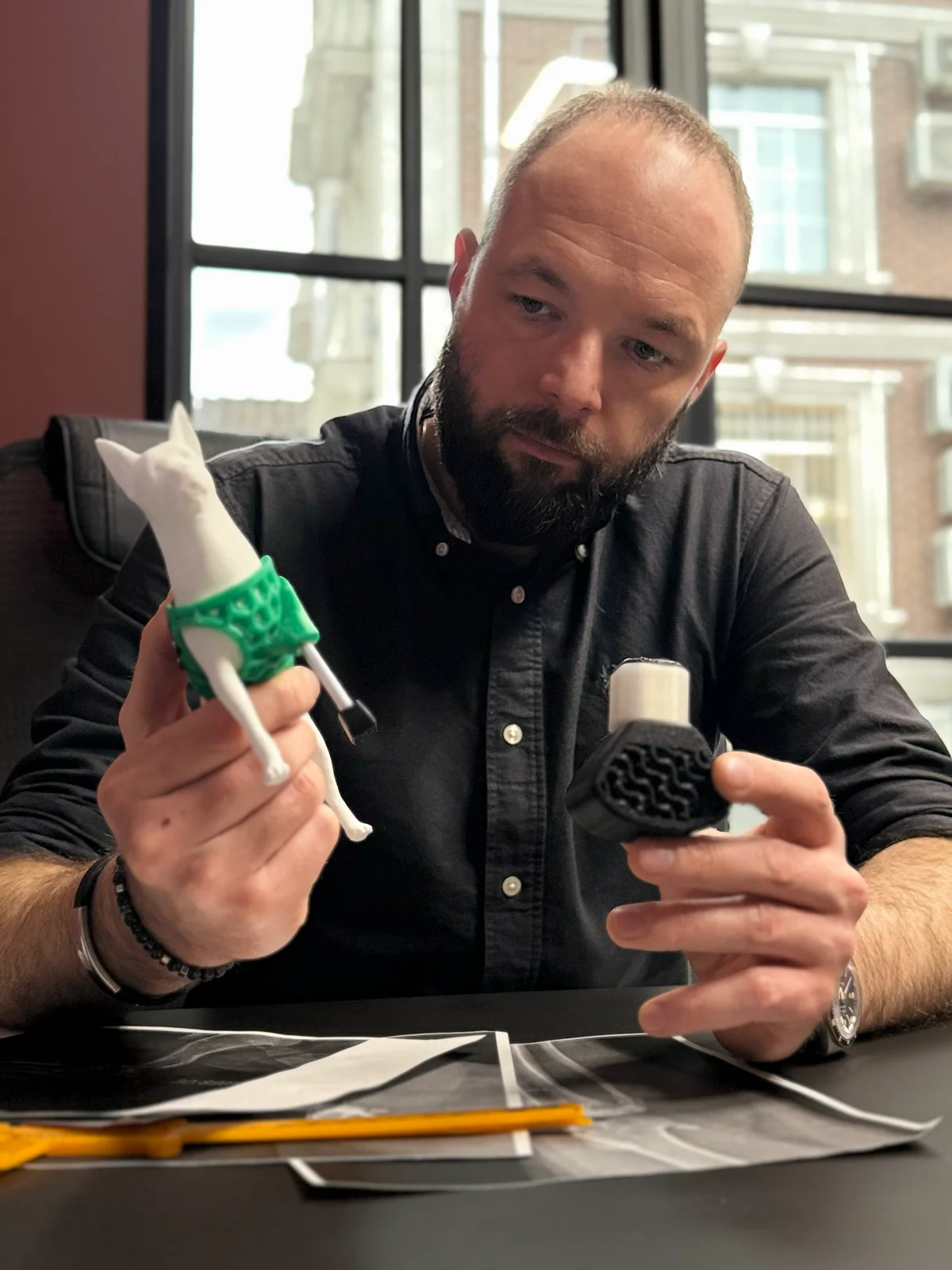
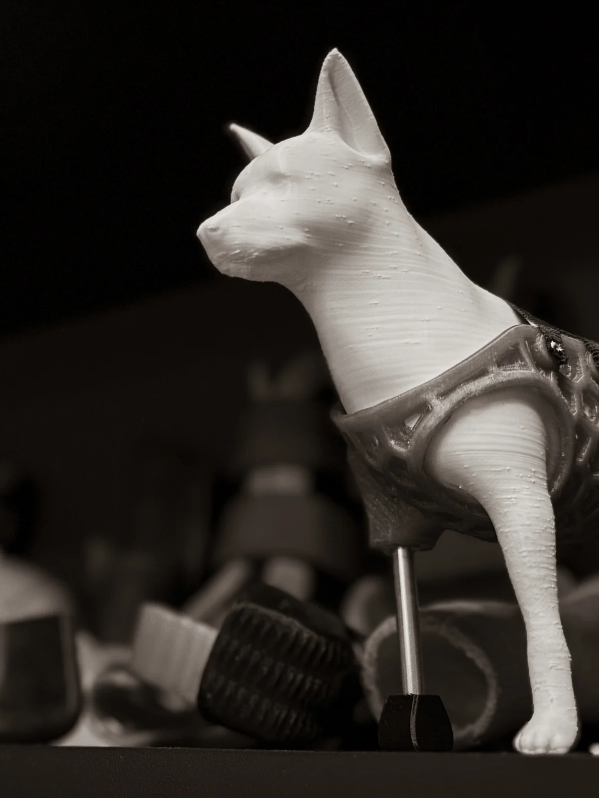
- creation of individual implants and prostheses - for complete anatomical correspondence;
- training - preparing the team for complex surgical interventions.
Advantages of 3D modeling: approach, fewer risks, more accurate positioning
- personalized approach to each patient;
- reducing the risk of complications and duration of surgery;
- precise positioning of implants and instruments;
- predicted treatment outcome;
- the ability to explain the operation to the owner using a visual 3D image.
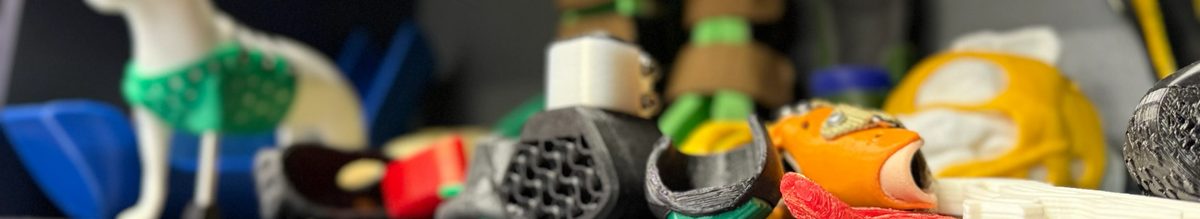
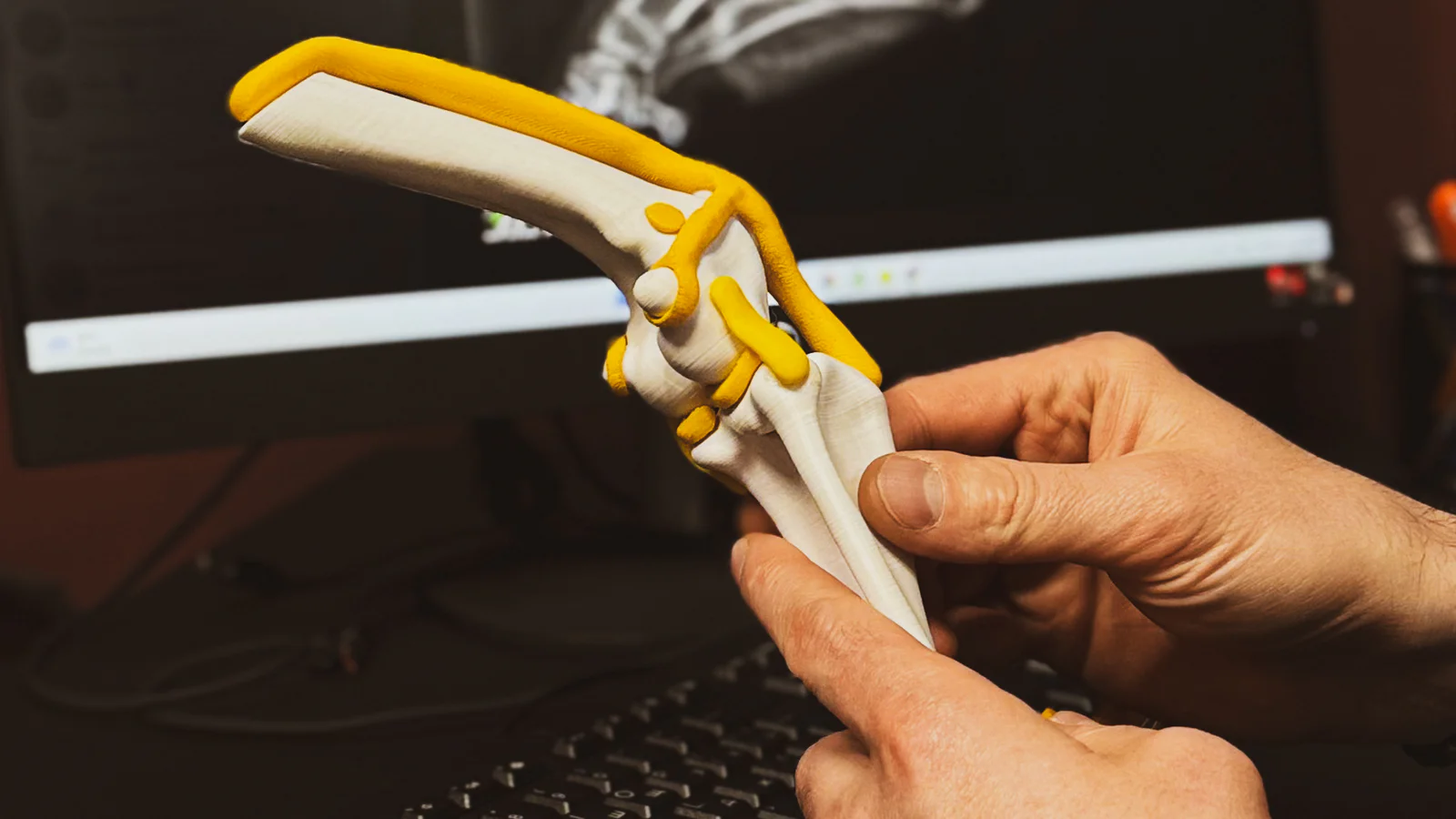
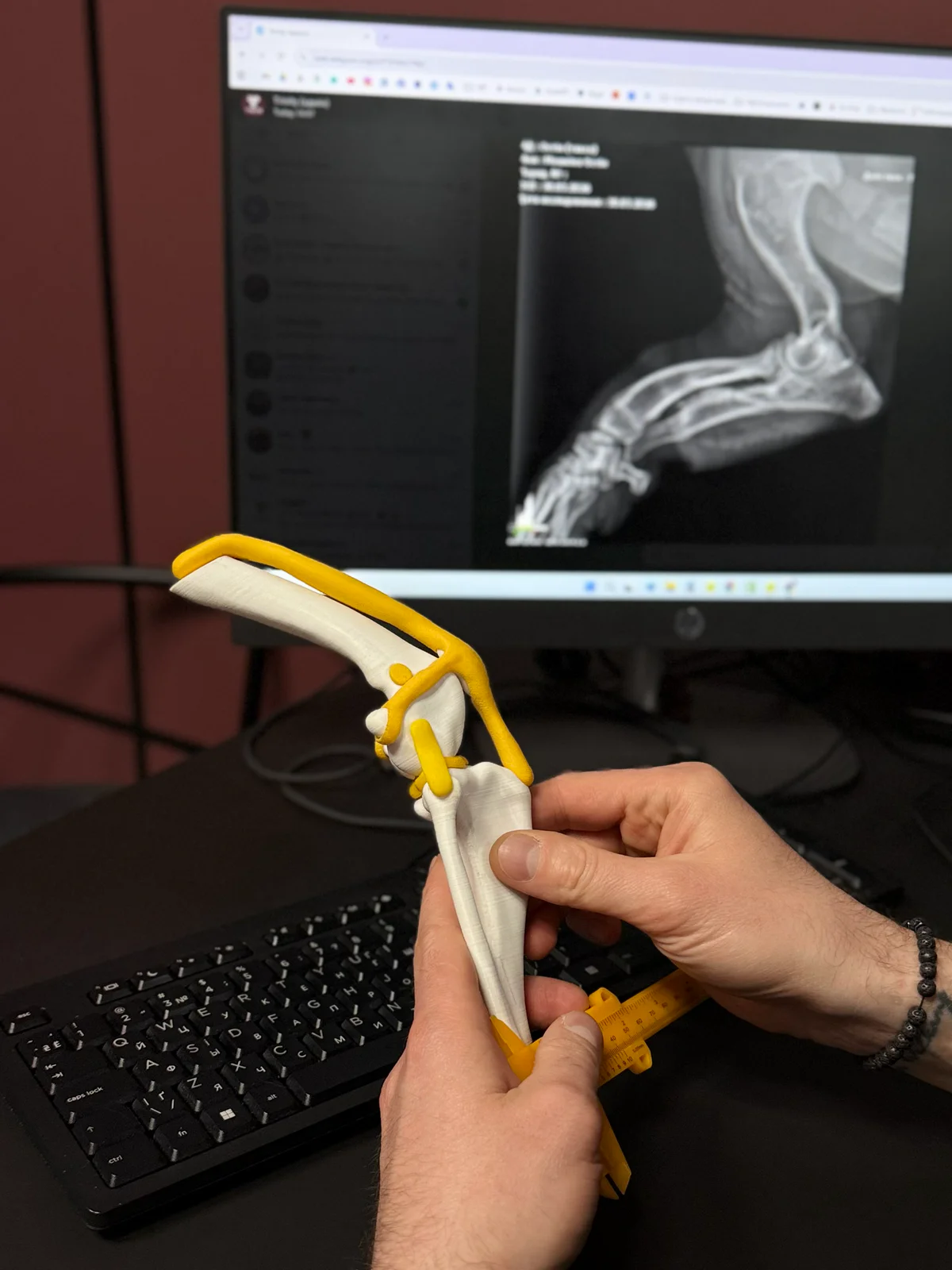
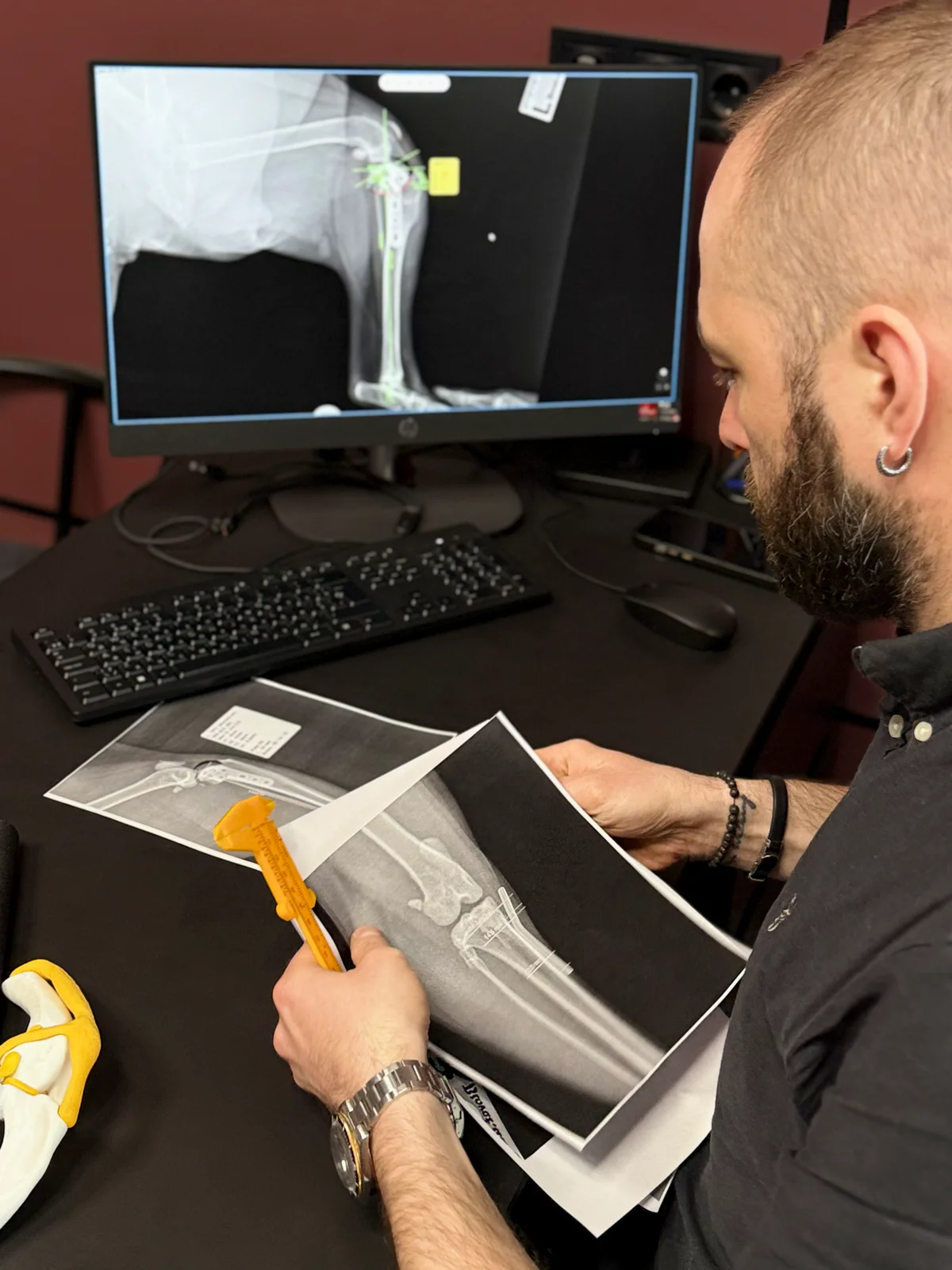
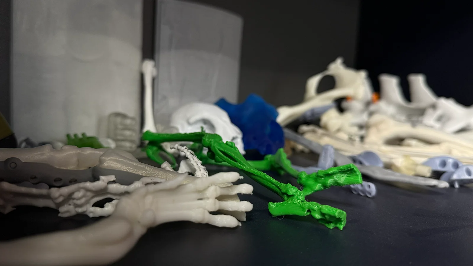
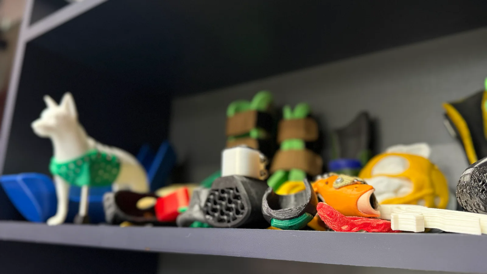
How 3D models are created: CT, MRI, digital reconstruction and 3D printing
3D models are created based on CT or MRI images of the animal using specialized software. The resulting models can be visualized in digital format or printed on a 3D printer for preliminary surgical planning. Trinity 3D modeling is precision that you can see. We make complex surgeries predictable and treatments effective and safe.
NEED A VETERINARY FOR YOUR PET?
Examination, diagnosis and treatment in one place
3D modeling in orthopedics, neurosurgery and oncology surgery: when it is especially useful
3D modeling in animal orthopedics helps the most when it is necessary to plan the correction of a complex fracture, deformity, or non-standard fixation. The doctor sees in advance exactly how the fragments are located, where it is better to draw a correction line, how to place a plate or screws. That is why 3D modeling of fractures in animals is not an «additional beautiful option,» but really reduces the number of unforeseen moments during the operation.
3D modeling is no less important in neurosurgery and oncology surgery of animals. When it comes to the skull, spine, tumors or critical anatomical areas, the surgeon must work as accurately as possible. Here, three-dimensional reconstruction of animals helps to assess the boundaries of the lesion and not act "by eye", but on the basis of a clear anatomical map.
Digital and Printed 3D Model: What is the Difference and How Does It Help the Surgeon?
In some cases, a digital model is sufficient. It allows you to scale the image, change the angle, assess the depth and relative location of structures. Such a digital 3D model of the animal is particularly useful for planning access, identifying risk areas and discussing tactics within the team. If the operation requires special precision, it may be advisable to 3D print an anatomical model of the animal.
A printed model allows you to literally hold the anatomy in your hands. This is important when you need to pre-fit a plate, check the shape of an implant, or understand how a complex area will look during an intervention. That is why 3D printing a model of an animal bone or other structure becomes a tool that helps reduce time in the operating room and increase the accuracy of movements.
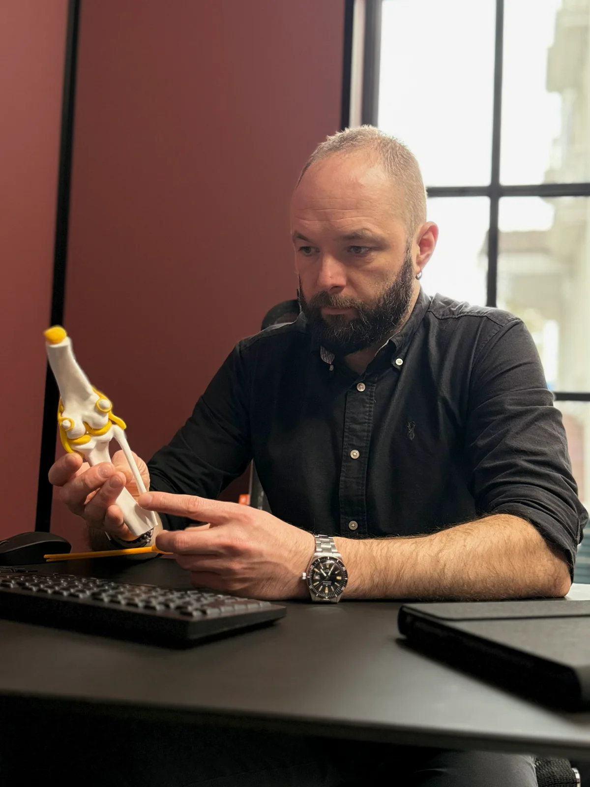
How a 3D model helps the owner understand the surgery plan
When an owner hears about a complex surgery, the biggest worry is the unknown. This is where a 3D model of the animal’s anatomy has another important advantage – it helps explain the problem clearly. The doctor can show exactly where the damage is, why a specific approach is needed, what exactly will be corrected, and what results are expected after the intervention.
This format of communication relieves some of the stress and allows you to make decisions not at random, but with understanding. It is important for us that a visual explanation of the operation to the owner is part of the respect for the person who entrusted us with their pet. When everything is visible, complex surgery ceases to be an abstraction and becomes a consistent, logical plan.
When 3D modeling is needed before a complex intervention: indications and possibilities
3D planning of an animal operation is especially useful when conventional images are not enough to fully understand the situation. This applies to complex fractures, non-standard deformations, tumors in hard-to-reach areas, operations on the spine, skull, or cases where it is necessary to combine several surgical tasks in one intervention. In such situations, preoperative 3D planning helps not to improvise during the operation, but to act according to a pre-thought-out route.
The technology is also particularly valuable when individual animal implants, anatomical guides or non-standard solutions are required. If we see that the conventional approach will not provide sufficient accuracy, it is surgical planning in animals with 3D reconstruction becomes a way to make future interventions more predictable and safer.
Advantages of 3D modeling at the Trinity Veterinary Hospital in Kyiv
At the Trinity Clinic in Kyiv, 3D modeling for animals combines diagnostics, surgical thinking, and modern digital technologies. We don’t use 3D just «for impression.» For us, it’s a tool that helps reduce operational risks, make accurate planning of animal surgery, and better prepare the team for a complex case.
Another advantage is that personalized approach in veterinary surgery really works here. We focus not on an average template, but on the anatomy of a specific patient. That is why the reduced risk of complications of the operation for the animal, more precise positioning of the instruments and a more predictable result become real, not declarative advantages.
How to book a 3D modeling consultation
To understand whether 3D modeling for animals is needed, it is important to briefly describe the clinical situation: what is the existing diagnosis, what studies have been performed, whether surgery is planned, whether there have been previous interventions. If you already have a CT or MRI scan, tell us about it right away, because it is CT scans for 3D modeling of an animal or MRI scans that most often become the basis for building a model.
If you are interested in a consultation on 3D modeling of an animal, an appointment for planning an intervention, or the cost of making a model, please contact the administrator for details. At the Trinity clinic in Kyiv, we will help you understand whether veterinary 3D modeling will be useful in your case, and how to properly prepare for the next stage of treatment.
READ MORE
Other branches

Do you need help?
Do you have questions or want to book your pet for an appointment?
Monday - Sunday: 24/7
Do you need help?
Do you have questions or want to book your pet for an appointment?
Monday - Sunday: 24/7
Consultation cost ophthalmologist
- All
- Inspection and Diagnostics
- Professional Sanitation
- Surgical Extraction
- Total Extraction
- Jaw Surgery
- Removal of Neoplasms
- Corrective Surgery
- Comprehensive Treatment (SONAT)
Inspection and Diagnostics
-
Initial dental examination
1,400 UAH.
-
Scheduled dental check-up
900 UAH.
-
Dental X-ray (one tooth)
600 UAH.
-
Detailed X-ray diagnostics
1,550 UAH.
Professional Sanitation
-
Ultrasonic sanitation of the oral cavity, 1st degree
2,600 UAH.
-
Ultrasonic sanitation of the oral cavity of the 2nd degree
3,100 UAH.
-
Ultrasonic sanitation of the oral cavity of the 3rd degree
3,600 UAH.
Surgical Extraction
-
Surgical tooth extraction of the 1st degree
700 UAH.
-
Surgical tooth extraction of the 2nd degree
850 UAH.
-
Surgical extraction of a 3rd degree tooth
1,050 UAH.
Total Extraction
-
Total tooth extraction of the 1st degree
5,600 UAH.
-
Total tooth extraction of the 2nd degree
7,600 UAH.
-
Total tooth extraction of the 3rd degree
9,600 UAH.
Jaw Surgery
-
Unilateral mandibulectomy
12,600 UAH.
-
Segmental mandibulectomy
12,600 UAH.
-
Rostral maxillectomy
15,600 UAH.
-
Caudal maxillectomy
18,600 UAH.
-
Total maxillectomy
20,600 UAH.
Removal of Neoplasms
-
Removal of a grade 1 soft tissue neoplasm
2,600 UAH.
-
Removal of a grade 2 soft tissue neoplasm
4,600 UAH.
-
Removal of a grade 3 soft tissue tumor
6,600 UAH.
Corrective Surgery
-
Surgical correction of a 1st degree cleft palate defect
2,600 UAH.
-
Surgical correction of a 2nd degree cleft palate defect
4,600 UAH.
-
Surgical correction of a 3rd degree cleft palate defect
8,600 UAH.
-
Surgical correction of grade 1 oronasal fistula
1,600 UAH.
-
Surgical correction of grade 2 oronasal fistula
2,600 UAH.
-
Surgical correction of grade 3 oronasal fistula
3,600 UAH.
Comprehensive Treatment (SONAT)
-
Comprehensive diagnostics and treatment according to the SONAT 1st degree protocol
4,000 UAH.
-
Comprehensive diagnostics and treatment according to the SONAT 2nd degree protocol
4,500 UAH.
-
Comprehensive diagnostics and treatment according to the SONAT 3rd degree protocol
5,000 UAH.
YOUR PET DESERVES THE BEST CARE
Entrust the health of your ponytail to professionals